JRC scientist contributed to the functionalisation and characterisation of silicon carbide nanoparticles (SiCNPs).
These particles have the promising potential to be used in cellular imaging applications for improved cancer diagnosis.
With the increase in life expectancy, cancer has become one of the most important health issues around the world.
Treatment efficacy correlates with early and accurate diagnosis. Thus continual improvement is necessary in diagnosis techniques, particularly in imaging.
Amongst the different methods, imaging techniques based on nonlinear optical (NLO) properties such as second-harmonic generation (SHG) and two photon excited fluorescence (TPEF) microscopies have been developed during this last decade.
However, most of the optical probes, both organic molecules and oxides nanoparticles used, suffers of different drawbacks namely low SHG signal and/or non-negligible toxicity.
In this study performed in collaboration with scientists from France, Switzerland and Ukraine a new class of NLO imaging probe based on SiC nanoparticles were developed, characterised and tested in vitro.
In particular, as a first step the surface of SiCNPs was chemically modified by using covalent reaction with 3-aminopropyltriethoxysilane (APTES) followed by grafting synthetic polyethylene glycol-folate (PEG-folate) molecules onto the amino-functionalized SiCNPs.
After each step of functionalisation, SiCNPs were characterised by zeta potential measurements and infrared spectroscopy.
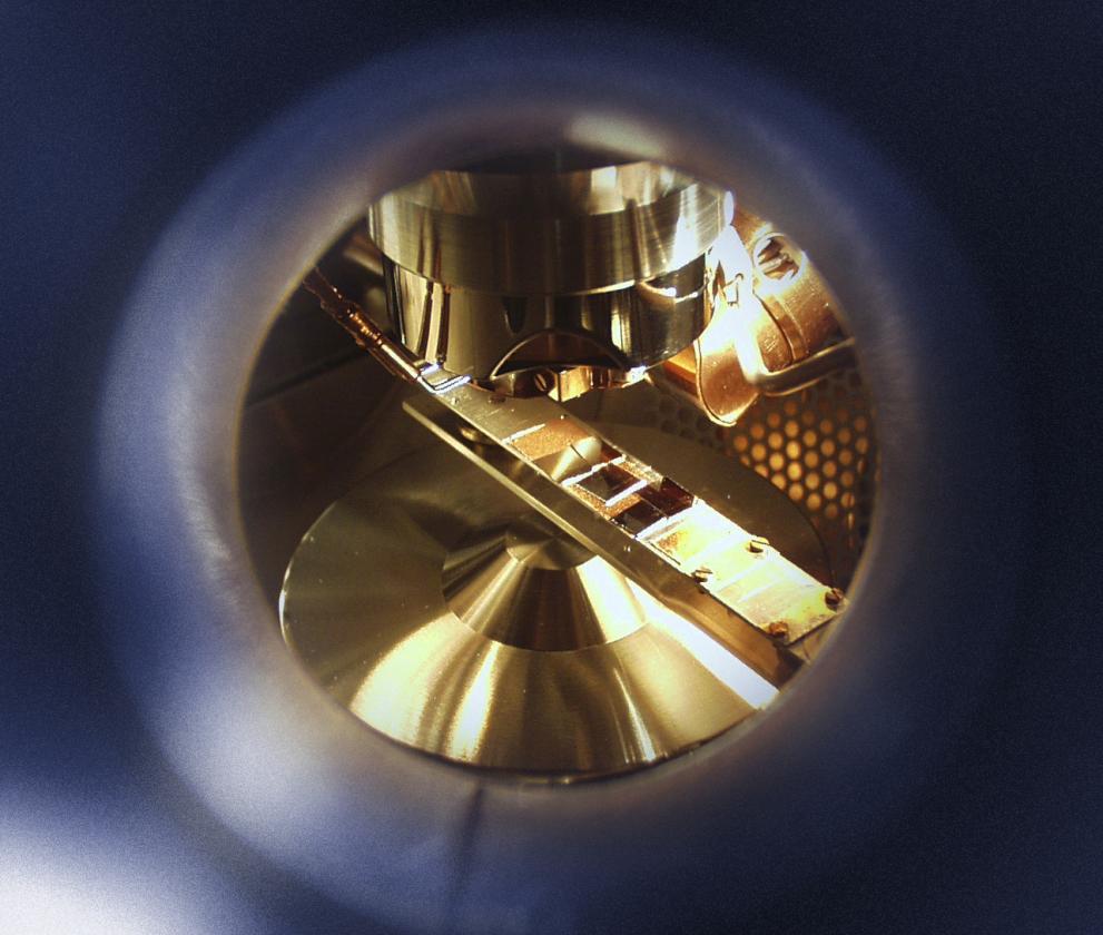
Amine surface density was quantified by a colorimetric titration method, whilst X-ray photoelectron spectroscopy (XPS) and time-of- flight secondary ion mass spectrometry (ToF-SIMS), performed at JRC, were used to identify the surface chemical composition and investigate the effect of different functionalization parameters.
Finally, the functionalised nanoparticles were tested in vitro using 3T3-L1 mouse fibroblasts and human hepatocarcinoma-derived HuH7 cells and the cancer-specific labelling efficiency of folate-modified SiCNPs was demonstrated by measurement of the SHG emitting cell area.

In conclusion, this study demonstrates that in respect to others NLO probes, SiCNPs offer the advantage of being a biocompatible material.
Moreover, because of the combination of a strong SHG signal that can be excited in the near-infrared region and the cancer-labelling specificity, PEG-folate-modified SiCNPs can be seriously considered as effective cancer-specific labels for multiphoton imaging of biopsy samples.
Read more in: M. Boksebeld et al.: 'Folate-modified silicon carbide nanoparticles as multiphoton imaging nanoprobes for cancer-cell specific labelling', RSC Adv. 7 (2017) 27361, doi:10.1039/C7RA03961A.
Related Content
Details
- Publication date
- 29 June 2017
